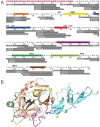
Thrombomodulin Binding Selects the Catalytically Active Form of Thrombin
- PMID: 26468766
- PMCID: PMC4697735
- DOI: 10.1021/acs.biochem.5b00825
Thrombomodulin Binding Selects the Catalytically Active Form of Thrombin
Abstract
Human α-thrombin is a serine protease with dual functions. Thrombin acts as a procoagulant, cleaving fibrinogen to make the fibrin clot, but when bound to thrombomodulin (TM), it acts as an anticoagulant, cleaving protein C. A minimal TM fragment consisting of the fourth, fifth, and most of the sixth EGF-like domain (TM456m) that has been prepared has much improved solubility, thrombin binding capacity, and anticoagulant activity versus those of previous TM456 constructs. In this work, we compare backbone amide exchange of human α-thrombin in three states: apo, D-Phe-Pro-Arg-chloromethylketone (PPACK)-bound, and TM456m-bound. Beyond causing a decreased level of amide exchange at their binding sites, TM and PPACK both cause a decreased level of amide exchange in other regions including the γ-loop and the adjacent N-terminus of the heavy chain. The decreased level of amide exchange in the N-terminus of the heavy chain is consistent with the historic model of activation of serine proteases, which involves insertion of this region into the β-barrel promoting the correct conformation of the catalytic residues. Contrary to crystal structures of thrombin, hydrogen-deuterium exchange mass spectrometry results suggest that the conformation of apo-thrombin does not yet have the N-terminus of the heavy chain properly inserted for optimal catalytic activity, and that binding of TM allosterically promotes the catalytically active conformation.
Figures


Similar articles
-
Identification of a thrombin-binding region in the sixth epidermal growth factor-like repeat of human thrombomodulin.Biochemistry. 2000 Aug 29;39(34):10365-72. doi: 10.1021/bi000715e. Biochemistry. 2000. PMID: 10956026
-
Allosteric changes in solvent accessibility observed in thrombin upon active site occupation.Biochemistry. 2004 May 11;43(18):5246-55. doi: 10.1021/bi0499718. Biochemistry. 2004. PMID: 15122890
-
The isomorphous structures of prethrombin2, hirugen-, and PPACK-thrombin: changes accompanying activation and exosite binding to thrombin.Protein Sci. 1994 Dec;3(12):2254-71. doi: 10.1002/pro.5560031211. Protein Sci. 1994. PMID: 7756983 Free PMC article.
-
Structure-function relationships of the thrombin-thrombomodulin interaction.Haemostasis. 1993 Mar;23 Suppl 1:183-93. doi: 10.1159/000216927. Haemostasis. 1993. PMID: 8388351 Review.
-
Thrombomodulin structure and function.Thromb Haemost. 1997 Jul;78(1):392-5. Thromb Haemost. 1997. PMID: 9198185 Review.
Cited by
-
Allosteric Modulation of Thrombin by Thrombomodulin: Insights from Logistic Regression and Statistical Analysis of Molecular Dynamics Simulations.ACS Omega. 2024 May 17;9(21):23086-23100. doi: 10.1021/acsomega.4c03375. eCollection 2024 May 28. ACS Omega. 2024. PMID: 38826540 Free PMC article.
-
The Light Chain Allosterically Enhances the Protease Activity of Murine Urokinase-Type Plasminogen Activator.Biochemistry. 2024 Jun 4;63(11):1434-1444. doi: 10.1021/acs.biochem.4c00071. Epub 2024 May 23. Biochemistry. 2024. PMID: 38780522 Free PMC article.
-
Simulations suggest double sodium binding induces unexpected conformational changes in thrombin.J Mol Model. 2022 Apr 13;28(5):120. doi: 10.1007/s00894-022-05076-0. J Mol Model. 2022. PMID: 35419655 Free PMC article.
-
How Thrombomodulin Enables W215A/E217A Thrombin to Cleave Protein C but Not Fibrinogen.Biochemistry. 2022 Jan 18;61(2):77-84. doi: 10.1021/acs.biochem.1c00635. Epub 2022 Jan 3. Biochemistry. 2022. PMID: 34978431 Free PMC article.
-
Advances in Hydrogen/Deuterium Exchange Mass Spectrometry and the Pursuit of Challenging Biological Systems.Chem Rev. 2022 Apr 27;122(8):7562-7623. doi: 10.1021/acs.chemrev.1c00279. Epub 2021 Sep 7. Chem Rev. 2022. PMID: 34493042 Free PMC article. Review.
References
-
- Furie B, Furie BC. The molecular basis of blood coagulation. Cell. 1988;53:505–518. - PubMed
-
- Bode W. Structure and interaction modes of thrombin. Blood Cells, Molecules, and Diseases. 2006;36:122–130. - PubMed
-
- Bode W. The transition of bovine trypsinogen to a trypsin-like state upon strong ligand binding: II. The binding of the pancreatic trypsin inhibitor and of isoleucine-valine and of sequentially related peptides to trypsinogen and to p-guanidinobenzoate trypsinogen. J Mol Biol. 1979;127:357–374. - PubMed
-
- Huber R, Bode W. Structural basis of the activation and action of trypsin. Acc Chem Res. 1978;11:114–122.
Publication types
MeSH terms
Substances
Grants and funding
LinkOut - more resources
Full Text Sources
Other Literature Sources

