Pigment Epithelium-Derived Factor Plays a Role in Alzheimer's Disease by Negatively Regulating Aβ42
- PMID: 29736859
- PMCID: PMC6095778
- DOI: 10.1007/s13311-018-0628-1
Pigment Epithelium-Derived Factor Plays a Role in Alzheimer's Disease by Negatively Regulating Aβ42
Abstract
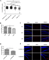
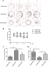
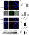
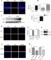
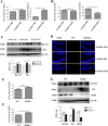
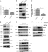
Alzheimer's disease (AD) is the most common cause of dementia. Pigment epithelium-derived factor (PEDF), a unique neurotrophic protein, decreases with aging. Previous reports have conflicted regarding whether the PEDF concentration is altered in AD patients. In addition, the effect of PEDF on AD has not been documented. Here, we tested serum samples of 31 AD patients and 271 normal controls. We found that compared to PEDF levels in young and middle-aged control subjects, PEDF levels were reduced in old-aged controls and even more so in AD patients. Furthermore, we verified that PEDF expression was much lower and amyloid β-protein (Aβ)42 expression was much higher in senescence-accelerated mouse prone 8 (SAMP8) strain mice than in senescence-accelerated mouse resistant 1 (SAMR1) control strain mice. Accordingly, high levels of Aβ42 were also observed in PEDF knockout (KO) mice. PEDF notably reduced cognitive impairment in the Morris water maze (MWM) and significantly downregulated Aβ42 in SAMP8 mice. Mechanistically, PEDF downregulated presenilin-1 (PS1) expression by inhibiting the c-Jun N-terminal kinase (JNK) pathway. Taken together, our findings demonstrate for the first time that PEDF negatively regulates Aβ42 and that PEDF deficiency with aging might play a crucial role in the development of AD.
Keywords: Alzheimer’s disease; Aβ42; Pigment epithelium-derived factor; Presenilin-1.
Figures

Similar articles
-
PEDF-derived peptide protects against Amyloid-β toxicity in vitro and prevents retinal dysfunction in rats.Exp Eye Res. 2024 May;242:109861. doi: 10.1016/j.exer.2024.109861. Epub 2024 Mar 23. Exp Eye Res. 2024. PMID: 38522635
-
Pigment epithelium-derived factor (PEDF) suppresses IL-1β-mediated c-Jun N-terminal kinase (JNK) activation to improve hepatocyte insulin signaling.Endocrinology. 2014 Apr;155(4):1373-85. doi: 10.1210/en.2013-1785. Epub 2014 Jan 23. Endocrinology. 2014. PMID: 24456163 Free PMC article.
-
Alzheimer's disease: Elevated pigment epithelium-derived factor in the cerebrospinal fluid is mostly of systemic origin.J Neurol Sci. 2017 Apr 15;375:123-128. doi: 10.1016/j.jns.2017.01.051. Epub 2017 Jan 17. J Neurol Sci. 2017. PMID: 28320113
-
The biological relevance of pigment epithelium-derived factor on the path from aging to age-related disease.Mech Ageing Dev. 2021 Jun;196:111478. doi: 10.1016/j.mad.2021.111478. Epub 2021 Apr 1. Mech Ageing Dev. 2021. PMID: 33812881 Review.
-
Pigment epithelium-derived factor regulation of neuronal and stem cell fate.Exp Cell Res. 2020 Apr 15;389(2):111891. doi: 10.1016/j.yexcr.2020.111891. Epub 2020 Feb 5. Exp Cell Res. 2020. PMID: 32035134 Review.
Cited by
-
Lycopene alleviates oxidative stress via the PI3K/Akt/Nrf2pathway in a cell model of Alzheimer's disease.PeerJ. 2020 Jun 8;8:e9308. doi: 10.7717/peerj.9308. eCollection 2020. PeerJ. 2020. PMID: 32551202 Free PMC article.
-
Roles of pigment epithelium-derived factor in exercise-induced suppression of senescence and its impact on lung pathology in mice.Aging (Albany NY). 2024 Jun 26;16(13):10670-10693. doi: 10.18632/aging.205976. Epub 2024 Jun 26. Aging (Albany NY). 2024. PMID: 38954512 Free PMC article.
-
Blood Proteome Profiling Reveals Biomarkers and Pathway Alterations in Fragile X PM at Risk for Developing FXTAS.Int J Mol Sci. 2023 Aug 30;24(17):13477. doi: 10.3390/ijms241713477. Int J Mol Sci. 2023. PMID: 37686279 Free PMC article.
-
Early Biomarkers of Neurodegenerative and Neurovascular Disorders in Diabetes.J Clin Med. 2020 Aug 30;9(9):2807. doi: 10.3390/jcm9092807. J Clin Med. 2020. PMID: 32872672 Free PMC article. Review.
-
The Role of PEDF in Reproductive Aging of the Ovary.Int J Mol Sci. 2022 Sep 8;23(18):10359. doi: 10.3390/ijms231810359. Int J Mol Sci. 2022. PMID: 36142276 Free PMC article.
References
Publication types
MeSH terms
Substances
Grants and funding
- 81471033/National Nature Science Foundation of China/International
- 81572342/National Nature Science Foundation of China/International
- 81600641/National Nature Science Foundation of China/International
- 81770808/National Nature Science Foundation of China/International
- 81370945/National Nature Science Foundation of China/International
- 81570871/National Nature Science Foundation of China/International
- 81570764/National Nature Science Foundation of China/International
- 2013ZX09102-053/National KeySci-Tech Special Project of China/International
- 2015GKS-355/National KeySci-Tech Special Project of China/International
- 2015A030311043/Key Project of Nature Science Foundation of Guangdong Province/International
- 2016A030311035/Key Project of Nature Science Foundation of Guangdong Province/International
- 2014A030313073;2015A030313029/Guangdong Natural Science Fund/International
- 2015A030313103/Guangdong Natural Science Fund/International
- 2014A020212023/Guangdong Natural Science Fund/International
- 2017A020215075/Guangdong Science Technology Project/International
- 2015B090903063/Guangdong Science Technology Project/International
- 13ykpy06/Initiate Research Funds for the Central Universities of China (Youth Program)/International
- 14ykpy05/Initiate Research Funds for the Central Universities of China (Youth Program)/International
- 16ykpy24/Initiate Research Funds for the Central Universities of China (Youth Program)/International
- 201707010084/Key Sci-tech Research Project of Guangzhou Municipality/International
- 201508020033/Key Sci-tech Research Project of Guangzhou Municipality/International
- 201610010186/Pearl River Nova Program of Guangzhou Municipality/International
- 201803010017/the Key Sci-tech Research Project of Guangzhou Municipality/International
LinkOut - more resources
Full Text Sources
Other Literature Sources
Medical
Research Materials
Miscellaneous

