Handheld Real-Time LED-Based Photoacoustic and Ultrasound Imaging System for Accurate Visualization of Clinical Metal Needles and Superficial Vasculature to Guide Minimally Invasive Procedures
- PMID: 29724014
- PMCID: PMC5982119
- DOI: 10.3390/s18051394
Handheld Real-Time LED-Based Photoacoustic and Ultrasound Imaging System for Accurate Visualization of Clinical Metal Needles and Superficial Vasculature to Guide Minimally Invasive Procedures
Abstract
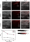
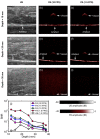
Ultrasound imaging is widely used to guide minimally invasive procedures, but the visualization of the invasive medical device and the procedure’s target is often challenging. Photoacoustic imaging has shown great promise for guiding minimally invasive procedures, but clinical translation of this technology has often been limited by bulky and expensive excitation sources. In this work, we demonstrate the feasibility of guiding minimally invasive procedures using a dual-mode photoacoustic and ultrasound imaging system with excitation from compact arrays of light-emitting diodes (LEDs) at 850 nm. Three validation experiments were performed. First, clinical metal needles inserted into biological tissue were imaged. Second, the imaging depth of the system was characterized using a blood-vessel-mimicking phantom. Third, the superficial vasculature in human volunteers was imaged. It was found that photoacoustic imaging enabled needle visualization with signal-to-noise ratios that were 1.2 to 2.2 times higher than those obtained with ultrasound imaging, over insertion angles of 26 to 51 degrees. With the blood vessel mimicking phantom, the maximum imaging depth was 38 mm. The superficial vasculature of a human middle finger and a human wrist were clearly visualized in real-time. We conclude that the LED-based system is promising for guiding minimally invasive procedures with peripheral tissue targets.
Keywords: LED; minimally invasive procedures; needle guidance; photoacoustic imaging; ultrasonography; vasculature.
Conflict of interest statement
The authors declare no conflict of interest.
Figures


Similar articles
-
Enhanced Photoacoustic Visualisation of Clinical Needles by Combining Interstitial and Extracorporeal Illumination of Elastomeric Nanocomposite Coatings.Sensors (Basel). 2022 Aug 25;22(17):6417. doi: 10.3390/s22176417. Sensors (Basel). 2022. PMID: 36080876 Free PMC article.
-
Improving needle visibility in LED-based photoacoustic imaging using deep learning with semi-synthetic datasets.Photoacoustics. 2022 Apr 7;26:100351. doi: 10.1016/j.pacs.2022.100351. eCollection 2022 Jun. Photoacoustics. 2022. PMID: 35495095 Free PMC article.
-
Performance characteristics of an interventional multispectral photoacoustic imaging system for guiding minimally invasive procedures.J Biomed Opt. 2015 Aug;20(8):86005. doi: 10.1117/1.JBO.20.8.086005. J Biomed Opt. 2015. PMID: 26263417 Free PMC article.
-
Towards Clinical Translation of LED-Based Photoacoustic Imaging: A Review.Sensors (Basel). 2020 Apr 27;20(9):2484. doi: 10.3390/s20092484. Sensors (Basel). 2020. PMID: 32349414 Free PMC article. Review.
-
Tutorial on phantoms for photoacoustic imaging applications.J Biomed Opt. 2024 Aug;29(8):080801. doi: 10.1117/1.JBO.29.8.080801. Epub 2024 Aug 14. J Biomed Opt. 2024. PMID: 39143981 Free PMC article. Review.
Cited by
-
Enhanced Photoacoustic Visualisation of Clinical Needles by Combining Interstitial and Extracorporeal Illumination of Elastomeric Nanocomposite Coatings.Sensors (Basel). 2022 Aug 25;22(17):6417. doi: 10.3390/s22176417. Sensors (Basel). 2022. PMID: 36080876 Free PMC article.
-
Fully Customized Photoacoustic System Using Doubly Q-Switched Nd:YAG Laser and Multiple Axes Stages for Laboratory Applications.Sensors (Basel). 2022 Mar 29;22(7):2621. doi: 10.3390/s22072621. Sensors (Basel). 2022. PMID: 35408235 Free PMC article.
-
Photoacoustic imaging in the second near-infrared window: a review.J Biomed Opt. 2019 Apr;24(4):1-20. doi: 10.1117/1.JBO.24.4.040901. J Biomed Opt. 2019. PMID: 30968648 Free PMC article. Review.
-
Handheld interventional ultrasound/photoacoustic puncture needle navigation based on deep learning segmentation.Biomed Opt Express. 2023 Oct 26;14(11):5979-5993. doi: 10.1364/BOE.504999. eCollection 2023 Nov 1. Biomed Opt Express. 2023. PMID: 38021141 Free PMC article.
-
Review of Low-Cost Photoacoustic Sensing and Imaging Based on Laser Diode and Light-Emitting Diode.Sensors (Basel). 2018 Jul 13;18(7):2264. doi: 10.3390/s18072264. Sensors (Basel). 2018. PMID: 30011842 Free PMC article. Review.
References
-
- Narouze S.N., editor. Atlas of Ultrasound-Guided Procedures in Interventional Pain Management. Springer Science & Business Media; New York, NY, USA: 2010.
-
- Rathmell J.P., Benzon H.T., Dreyfuss P., Huntoon M., Wallace M., Baker R., Riew K.D., Rosenquist R.W., Aprill C., Rost N.S., et al. Safeguards to Prevent Neurologic Complications after Epidural Steroid Injections: Consensus Opinions from a Multidisciplinary Working Group and National Organizations. Surv. Anesthesiol. 2016;60:85–86. doi: 10.1097/01.sa.0000480641.01172.93. - DOI - PubMed
MeSH terms
Substances
LinkOut - more resources
Full Text Sources
Other Literature Sources

