High-Resolution Analysis of Antibodies to Post-Translational Modifications Using Peptide Nanosensor Microarrays
- PMID: 27636738
- PMCID: PMC5367622
- DOI: 10.1021/acsnano.6b03786
High-Resolution Analysis of Antibodies to Post-Translational Modifications Using Peptide Nanosensor Microarrays
Abstract
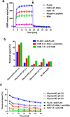
Autoantibodies are a hallmark of autoimmune diseases such as lupus and have the potential to be used as biomarkers for diverse diseases, including immunodeficiency, infectious disease, and cancer. More precise detection of antibodies to specific targets is needed to improve diagnosis of such diseases. Here, we report the development of reusable peptide microarrays, based on giant magnetoresistive (GMR) nanosensors optimized for sensitively detecting magnetic nanoparticle labels, for the detection of antibodies with a resolution of a single post-translationally modified amino acid. We have also developed a chemical regeneration scheme to perform multiplex assays with a high level of reproducibility, resulting in greatly reduced experimental costs. In addition, we show that peptides synthesized directly on the nanosensors are approximately two times more sensitive than directly spotted peptides. Reusable peptide nanosensor microarrays enable precise detection of autoantibodies with high resolution and sensitivity and show promise for investigating antibody-mediated immune responses to autoantigens, vaccines, and pathogen-derived antigens as well as other fundamental peptide-protein interactions.
Keywords: autoantibody; giant magnetoresistance; lupus; nanosensors; peptide microarray; regeneration.
Figures


Similar articles
-
Autoantigen microarrays for multiplex characterization of autoantibody responses.Nat Med. 2002 Mar;8(3):295-301. doi: 10.1038/nm0302-295. Nat Med. 2002. PMID: 11875502
-
Multiplex giant magnetoresistive biosensor microarrays identify interferon-associated autoantibodies in systemic lupus erythematosus.Sci Rep. 2016 Jun 9;6:27623. doi: 10.1038/srep27623. Sci Rep. 2016. PMID: 27279139 Free PMC article.
-
The use of post-translationally modified peptides for detection of biomarkers of immune-mediated diseases.J Pept Sci. 2009 Oct;15(10):621-8. doi: 10.1002/psc.1166. J Pept Sci. 2009. PMID: 19714713 Review.
-
Mapping epitopes of U1-70K autoantibodies at single-amino acid resolution.Autoimmunity. 2015;48(8):513-23. doi: 10.3109/08916934.2015.1077233. Epub 2015 Aug 31. Autoimmunity. 2015. PMID: 26333287 Free PMC article.
-
Applications of Peptide Microarrays in Autoantibody, Infection, and Cancer Detection.Methods Mol Biol. 2023;2578:1-15. doi: 10.1007/978-1-0716-2732-7_1. Methods Mol Biol. 2023. PMID: 36152276 Review.
Cited by
-
Improved detection of prostate cancer using a magneto-nanosensor assay for serum circulating autoantibodies.PLoS One. 2019 Aug 12;14(8):e0221051. doi: 10.1371/journal.pone.0221051. eCollection 2019. PLoS One. 2019. PMID: 31404106 Free PMC article. Clinical Trial.
-
Longitudinal Multiplexed Measurement of Quantitative Proteomic Signatures in Mouse Lymphoma Models Using Magneto-Nanosensors.Theranostics. 2018 Feb 3;8(5):1389-1398. doi: 10.7150/thno.20706. eCollection 2018. Theranostics. 2018. PMID: 29507628 Free PMC article.
-
Magnetic supercluster particles for highly sensitive magnetic biosensing of proteins.Mikrochim Acta. 2022 Jun 14;189(7):256. doi: 10.1007/s00604-022-05354-x. Mikrochim Acta. 2022. PMID: 35697882 Free PMC article.
-
Design and Fabrication of Full Wheatstone-Bridge-Based Angular GMR Sensors.Sensors (Basel). 2018 Jun 5;18(6):1832. doi: 10.3390/s18061832. Sensors (Basel). 2018. PMID: 29874825 Free PMC article.
-
Longitudinal Monitoring of Antibody Responses against Tumor Cells Using Magneto-nanosensors with a Nanoliter of Blood.Nano Lett. 2017 Nov 8;17(11):6644-6652. doi: 10.1021/acs.nanolett.7b02591. Epub 2017 Oct 20. Nano Lett. 2017. PMID: 28990786 Free PMC article.
References
-
- Yaniv G, Twig G, Shor DB, Furer A, Sherer Y, Mozes O, Komisar O, Slonimsky E, Kiang E, Lotan E, Welt M, Maraj I, Shina A, Amital H, Shoenfeld Y. A Volcanic Explosion of Autoantibodies in Systemic Lupus Erythematosus: a Diversity of 180 Different Antibodies Found in SLE Patients. Autoimmun. Rev. 2015;14:75–79. - PubMed
-
- Arbuckle MR, McClain MT, Rubertone MV, Scofield RH, Dennis GJ, James JA, Harley JB. Development of Autoantibodies before the Clinical Onset of Systemic Lupus Erythematosus. N. Engl. J. Med. 2003;349:1526–1533. - PubMed
-
- Nielen MM, van Schaardenburg D, Reesink HW, van de Stadt RJ, van der Horst-Bruinsma IE, de Koning MH, Habibuw MR, Vandenbroucke JP, Dijkmans BA. Specific Autoantibodies Precede the Symptoms of Rheumatoid Arthritis: a Study of Serial Measurements in Blood Donors. Arthritis Rheum. 2004;50:380–386. - PubMed
Publication types
MeSH terms
Substances
Grants and funding
LinkOut - more resources
Full Text Sources
Other Literature Sources
Research Materials

