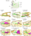
Palaeoneurological clues to the evolution of defining mammalian soft tissue traits
- PMID: 27157809
- PMCID: PMC4860582
- DOI: 10.1038/srep25604
Palaeoneurological clues to the evolution of defining mammalian soft tissue traits
Abstract
A rich fossil record chronicles the distant origins of mammals, but the evolution of defining soft tissue characters of extant mammals, such as mammary glands and hairs is difficult to interpret because soft tissue does not readily fossilize. As many soft tissue features are derived from dermic structures, their evolution is linked to that of the nervous syutem, and palaeoneurology offers opportunities to find bony correlates of these soft tissue features. Here, a CT scan study of 29 fossil skulls shows that non-mammaliaform Prozostrodontia display a retracted, fully ossified, and non-ramified infraorbital canal for the infraorbital nerve, unlike more basal therapsids. The presence of a true infraorbital canal in Prozostrodontia suggests that a motile rhinarium and maxillary vibrissae were present. Also the complete ossification of the parietal fontanelle (resulting in the loss of the parietal foramen) and the development of the cerebellum in Probainognathia may be pleiotropically linked to the appearance of mammary glands and having body hair coverage since these traits are all controlled by the same homeogene, Msx2, in mice. These suggest that defining soft tissue characters of mammals were already present in their forerunners some 240 to 246 mya.
Conflict of interest statement
The authors declare no competing interests.
Figures


Similar articles
-
The relationship between the infraorbital foramen, infraorbital nerve, and maxillary mechanoreception: implications for interpreting the paleoecology of fossil mammals based on infraorbital foramen size.Anat Rec (Hoboken). 2008 Oct;291(10):1221-6. doi: 10.1002/ar.20742. Anat Rec (Hoboken). 2008. PMID: 18780305
-
Craniodental anatomy in Permian-Jurassic Cynodontia and Mammaliaformes (Synapsida, Therapsida) as a gateway to defining mammalian soft tissue and behavioural traits.Philos Trans R Soc Lond B Biol Sci. 2023 Jul 3;378(1880):20220084. doi: 10.1098/rstb.2022.0084. Epub 2023 May 15. Philos Trans R Soc Lond B Biol Sci. 2023. PMID: 37183903 Free PMC article. Review.
-
A comparative analysis of infraorbital foramen size in Paleogene euarchontans.J Hum Evol. 2017 Apr;105:57-68. doi: 10.1016/j.jhevol.2017.01.017. Epub 2017 Mar 18. J Hum Evol. 2017. PMID: 28366200
-
The postcranial anatomy of Brasilodon quadrangularis and the acquisition of mammaliaform traits among non-mammaliaform cynodonts.PLoS One. 2019 May 10;14(5):e0216672. doi: 10.1371/journal.pone.0216672. eCollection 2019. PLoS One. 2019. PMID: 31075140 Free PMC article.
-
Avian palaeoneurology: Reflections on the eve of its 200th anniversary.J Anat. 2020 Jun;236(6):965-979. doi: 10.1111/joa.13160. Epub 2020 Jan 30. J Anat. 2020. PMID: 31999834 Free PMC article. Review.
Cited by
-
Oxygen isotopes suggest elevated thermometabolism within multiple Permo-Triassic therapsid clades.Elife. 2017 Jul 18;6:e28589. doi: 10.7554/eLife.28589. Elife. 2017. PMID: 28716184 Free PMC article.
-
Reptile-like physiology in Early Jurassic stem-mammals.Nat Commun. 2020 Oct 12;11(1):5121. doi: 10.1038/s41467-020-18898-4. Nat Commun. 2020. PMID: 33046697 Free PMC article.
-
Co-Evolution of Breast Milk Lipid Signaling and Thermogenic Adipose Tissue.Biomolecules. 2021 Nov 16;11(11):1705. doi: 10.3390/biom11111705. Biomolecules. 2021. PMID: 34827703 Free PMC article. Review.
-
New evidence from high-resolution computed microtomography of Triassic stem-mammal skulls from South America enhances discussions on turbinates before the origin of Mammaliaformes.Sci Rep. 2024 Jun 15;14(1):13817. doi: 10.1038/s41598-024-64434-5. Sci Rep. 2024. PMID: 38879680 Free PMC article.
-
The mystery of a missing bone: revealing the orbitosphenoid in basal Epicynodontia (Cynodontia, Therapsida) through computed tomography.Naturwissenschaften. 2017 Aug;104(7-8):66. doi: 10.1007/s00114-017-1487-z. Epub 2017 Jul 18. Naturwissenschaften. 2017. PMID: 28721557
References
-
- Martin T. et al.. A Cretaceous eutriconodont and integument evolution in early mammals. Nature 526, 380–384 (2015). - PubMed
-
- Oftedal O. T. The mammary gland and its origin during synapsid evolution. J Mammary Gland Biol Neoplasia 7, 225–252 (2002). - PubMed
-
- Lefèvre C. M., Sharp J. A. & Nicholas K. R. Evolution of Lactation: Ancient Origin and Extreme Adaptations of the Lactation System. Annu Rev Genomics Hum Genet 11, 219–38 (2010). - PubMed
-
- Satokata I. et al.. Msx2 deficiency in mice causes pleiotropic defects in bone growth and ectodermal organ formation. Nat Genet 24, 391–395 (2000). - PubMed
-
- Ji Q., Luo Z.-X., Yuan C.-X. & Tabrum A. R. A swimming mammaliaform from the Middle Jurassic and ecomorphological diversification of early mammals. Science 311(5764), 1123–1127 (2006). - PubMed
Publication types
MeSH terms
LinkOut - more resources
Full Text Sources
Other Literature Sources
Research Materials

