Actin and Actin-Binding Proteins - PubMed Skip to main page content
U.S. flag

An official website of the United States government

Dot gov

The .gov means it’s official.
Federal government websites often end in .gov or .mil. Before sharing sensitive information, make sure you’re on a federal government site.

Https

The site is secure.
The https:// ensures that you are connecting to the official website and that any information you provide is encrypted and transmitted securely.

Access keys NCBI Homepage MyNCBI Homepage Main Content Main Navigation
Review
. 2016 Aug 1;8(8):a018226.
doi: 10.1101/cshperspect.a018226.

Actin and Actin-Binding Proteins

Affiliations
Review

Actin and Actin-Binding Proteins

Thomas D Pollard. Cold Spring Harb Perspect Biol. .

Abstract

Organisms from all domains of life depend on filaments of the protein actin to provide structure and to support internal movements. Many eukaryotic cells use forces produced by actin polymerization for their motility, and myosin motor proteins use ATP hydrolysis to produce force on actin filaments. Actin polymerizes spontaneously, followed by hydrolysis of a bound adenosine triphosphate (ATP). Dissociation of the γ-phosphate prepares the polymer for disassembly. This review provides an overview of the properties of actin and shows how dozens of proteins control both the assembly and disassembly of actin filaments. These players catalyze nucleotide exchange on actin monomers, initiate polymerization, promote phosphate dissociation, cap the ends of polymers, cross-link filaments to each other and other cellular components, and sever filaments.


Figures

Figure 1.
Figure 1.
Structures of the actin molecule and actin filament. (A) Ribbon diagram of the actin molecule with space-filling ATP (protein data bank [PDB]: 1ATN). N, amino terminus; C, carboxyl terminus. Numbers 1, 2, 3, and 4 label the four subdomains. (B) Space-filling model of actin showing the nucleotide-binding cleft with ATP in situ and barbed-end groove. (C) Reconstruction of the actin filament from cryo-electron micrographs. The labels are single-letter abbreviations for selected amino acids. (D) Cartoon of the actin filament showing the position of the pointed and barbed ends. (A,B, Reprinted, with permission, from Pollard and Earnshaw 2007; C, reprinted, with permission from Macmillan Publishers Ltd., from Fujii et al. 2010; D, adapted, with permission, from Pollard and Earnshaw 2007.)
Figure 2.
Figure 2.
Actin polymerization. (A) Electron micrograph of a negatively stained actin filament. A seed was first decorated with myosin heads and then allowed to grow bare extensions. Elongation was faster at the barbed end than at the pointed end. (B) Diagram showing the rate constants for actin association and dissociation at the two ends of an actin filament. The pointed end is at the top and the barbed end is at the bottom. Unit of association rate constants, µm−1 sec−1; unit of dissociation rate constants, sec−1. The K values are the ratios of dissociation rate constants to association rate constants, the critical concentrations for each of the four reactions. The horizontal arrows indicate the exchange of adenosine diphosphate (ADP) for ATP. (C) Time course of spontaneous polymerization of Mg-ATP–actin monomers. The solid line is the polymer concentration measured by the fluorescence of pyrene-labeled actin. The initial lag comes from slow spontaneous nucleation. The reaction reaches a steady state when the free actin monomer concentration reaches the overall critical concentration. Filled circles are the extent of hydrolysis of the bound ATP, which lags behind polymerization by a few seconds. (D) Mechanism of nucleation, showing monomers, a dimer, a trimer, and a filament, with estimates of the rate constants for each step. Unit of association rate constants, µm−1 sec−1; unit of dissociation rate constants, sec−1. (A,B,D, Adapted, with permission, from Pollard and Earnshaw 2007; C, reprinted from Pollard and Weeds 1984.)
Figure 3.
Figure 3.
Nucleotide reactions of actin. The cartoon shows an actin filament that has grown from both ends of an ADP–actin seed. Over time, ATP bound to the polymerized subunits is hydrolyzed randomly to ADP and phosphate (Pi), followed by slow dissociation of the phosphate, leaving ADP–actin. ADP dissociates from ADP–actin monomers and is rapidly replaced by ATP. Profilin speeds ADP dissociation. (Adapted, with permission, from Pollard and Earnshaw 2007.)
Figure 4.
Figure 4.
Overview of families of actin-binding proteins, including monomer binding, polymerases such as formins, capping proteins, severing proteins, cross-linking proteins, and branching protein Arp2/3 complex. Filaments can anneal end to end, but no proteins are known to facilitate this reaction. The drawing does not include tropomyosin and myosin motors, which bind to the sides of filaments. (Adapted, with permission, from Pollard and Earnshaw 2007.)
Figure 5.
Figure 5.
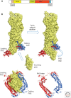
Proteins that bind actin monomers. Space-filling models of the actin monomer with ribbon diagrams of bound proteins. This is the standard view of actin (see Fig. 1), with the ATP-binding cleft at the top and the barbed-end groove at the bottom. (A) The WH2 helix binds in the barbed-end groove (PDB: 3M1F, from Vibrio parahaemolyticus Vopl). (B) Thymosin-β4 helices bind in both the barbed-end groove and across the pointed-end cleft (PDB: 4PL7). (C) Profilin can bind simultaneously to the barbed end of actin and to polyproline helices such as that from vasodilator-stimulated phosphoprotein (VASP), shown here as a red stick figure (PDB: 2PBD). (D) The carboxy-terminal cofilin domain from twinfilin binds on the barbed end of the actin molecule (PDB: 3DAW).
Figure 6.
Figure 6.
The Arp2/3 complex. (A) Ribbon diagram showing the two actin-related proteins (Arps) and the five novel subunits. Subdomains 1 and 2 of Arp2 were disordered in this structure (PDB: 1K8K). (B) Model of the branch junction from reconstructions of electron micrographs (Rouiller et al. 2008). The Arp2/3 complex anchors a branch represented by daughter subunits D1 and D2 on the side of the mother filament. The numbers indicate the subdomains of Arp2 (red) and Arp3 (orange). (C) Model (Padrick et al. 2011) for the interaction of Arp2/3 complex with two verprolin-cofilin-acidic (VCA) motifs (black), each with an actin subunit bound to the V (WH2) motif (Chereau et al. 2005). The location of the A motif on Arp3 was determined by X-ray crystallography (Ti et al. 2011). Other aspects of the model were inferred from binding and cross-linking experiments (Padrick et al. 2011). (A, Reprinted, with permission, from Pollard and Earnshaw 2007; B, reprinted from Rouiller et al. 2008; C, adapted, with permission, from Padrick et al. 2011.)
Figure 7.
Figure 7.
Structure and role of formins. (A) Domain organization of a diaphanous (Dia)-type formin. DAD, diaphanous autoinhibitory domain; DID, DAD-interacting domain; GBD, GTPase-binding domain; FH1, proline-rich formin homology 1 domain; FH2, formin homology 2 domain. (B) Model of the actin filament elongation mechanism, with actin subunits shown by a space-filling model and the formin Bni1 FH2 dimer shown as red and blue ribbon diagrams. The model is based on the crystal structure (Otomo et al. 2005) docked on the barbed end of the actin filament model from fiber diffraction (Oda et al. 2009) and refined by molecular-dynamics simulation (Baker et al. 2015). An actin subunit binds from solution, creating a binding site for the trailing FH2 domain (blue) to “step” toward the barbed end. The lower diagrams show end-on views, including (left) the actin filament and (right) the FH2 dimer alone.
Figure 8.
Figure 8.
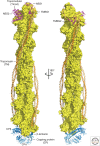
Capping of the two ends of the actin filament. Depicted is a space-filling model of a short filament with two laterally aligned tropomyosin molecules (orange), terminating with tropomodulin at the filament pointed end (magenta) (Rao et al. 2014) and heterodimeric capping protein (CP; cyan and blue) at the filament barbed end (Urnavicius et al. 2015). (Figure prepared by Roberto Dominguez of the University of Pennsylvania.)
Figure 9.
Figure 9.
Cross-linking proteins. Illustrations depicting the size, domains, and organization of the actin filament cross-linking proteins fimbrin, α-actinin, and filamin (ABP). The different patterns of distribution of the actin-binding domains lend the proteins distinct cross-linking capabilities. ABD, actin-binding domain; EF hand, calcium-binding motif. (Adapted from Pollard and Earnshaw 2007, with permission from Elsevier.)

Similar articles

Cited by

References

    1. Alberts AS. 2001. Identification of a carboxyl-terminal diaphanous-related formin homology protein autoregulatory domain. J Biol Chem 276: 2824–2830. - PubMed
    1. Andrianantoandro E, Pollard TD. 2006. Mechanism of actin filament turnover by severing and nucleation at different concentrations of ADF/cofilin. Mol Cell 24: 13–23. - PubMed
    1. Archer SJ, Vinson VK, Pollard TD, Torchia DA. 1994. Elucidation of the poly-l-proline binding site in Acanthamoeba profilin-I by NMR spectroscopy. FEBS Lett 337: 145–151. - PubMed
    1. Baker JL, Courtemanche N, Parton DL, McCullagh M, Pollard TD, Voth GA. 2015. Electrostatic interactions between the Bni1p formin FH2 domain and actin influence actin filament nucleation. Structure 23: 68–79. - PMC - PubMed
    1. Bamburg JR, Harris HE, Weeds AG. 1980. Partial purification and characterization of an actin depolymerizing factor from brain. FEBS Lett 121: 178–182. - PubMed

Publication types

LinkOut - more resources