Multicenter imaging outcomes study of The Cancer Genome Atlas glioblastoma patient cohort: imaging predictors of overall and progression-free survival
- PMID: 26203066
- PMCID: PMC4648306
- DOI: 10.1093/neuonc/nov117
Multicenter imaging outcomes study of The Cancer Genome Atlas glioblastoma patient cohort: imaging predictors of overall and progression-free survival
Abstract
Background: Despite an aggressive therapeutic approach, the prognosis for most patients with glioblastoma (GBM) remains poor. The aim of this study was to determine the significance of preoperative MRI variables, both quantitative and qualitative, with regard to overall and progression-free survival in GBM.
Methods: We retrospectively identified 94 untreated GBM patients from the Cancer Imaging Archive who had pretreatment MRI and corresponding patient outcomes and clinical information in The Cancer Genome Atlas. Qualitative imaging assessments were based on the Visually Accessible Rembrandt Images feature-set criteria. Volumetric parameters were obtained of the specific tumor components: contrast enhancement, necrosis, and edema/invasion. Cox regression was used to assess prognostic and survival significance of each image.
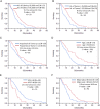
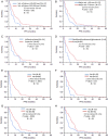
Results: Univariable Cox regression analysis demonstrated 10 imaging features and 2 clinical variables to be significantly associated with overall survival. Multivariable Cox regression analysis showed that tumor-enhancing volume (P = .03) and eloquent brain involvement (P < .001) were independent prognostic indicators of overall survival. In the multivariable Cox analysis of the volumetric features, the edema/invasion volume of more than 85 000 mm(3) and the proportion of enhancing tumor were significantly correlated with higher mortality (Ps = .004 and .003, respectively).
Conclusions: Preoperative MRI parameters have a significant prognostic role in predicting survival in patients with GBM, thus making them useful for patient stratification and endpoint biomarkers in clinical trials.
Keywords: TCGA; glioblastoma; imaging; overall survival; progression free survival.
© The Author(s) 2015. Published by Oxford University Press on behalf of the Society for Neuro-Oncology. All rights reserved. For permissions, please e-mail: journals.permissions@oup.com.
Figures


Similar articles
-
Algorithmic three-dimensional analysis of tumor shape in MRI improves prognosis of survival in glioblastoma: a multi-institutional study.J Neurooncol. 2017 Mar;132(1):55-62. doi: 10.1007/s11060-016-2359-7. Epub 2017 Jan 10. J Neurooncol. 2017. PMID: 28074320
-
Texture Feature Ratios from Relative CBV Maps of Perfusion MRI Are Associated with Patient Survival in Glioblastoma.AJNR Am J Neuroradiol. 2016 Jan;37(1):37-43. doi: 10.3174/ajnr.A4534. Epub 2015 Oct 15. AJNR Am J Neuroradiol. 2016. PMID: 26471746 Free PMC article.
-
Semantic imaging features predict disease progression and survival in glioblastoma multiforme patients.Strahlenther Onkol. 2018 Jun;194(6):580-590. doi: 10.1007/s00066-018-1276-4. Epub 2018 Feb 13. Strahlenther Onkol. 2018. PMID: 29442128 English.
-
Prior malignancies in patients harboring glioblastoma: an institutional case-study of 2164 patients.J Neurooncol. 2017 Sep;134(2):245-251. doi: 10.1007/s11060-017-2512-y. Epub 2017 May 27. J Neurooncol. 2017. PMID: 28551847 Review.
-
Noninvasive Glioblastoma Testing: Multimodal Approach to Monitoring and Predicting Treatment Response.Dis Markers. 2018 Jan 17;2018:2908609. doi: 10.1155/2018/2908609. eCollection 2018. Dis Markers. 2018. PMID: 29581794 Free PMC article. Review.
Cited by
-
Prognostic value of subventricular zone involvement in relation to tumor volumes defined by fused MRI and O-(2-[18F]fluoroethyl)-L-tyrosine (FET) PET imaging in glioblastoma multiforme.Radiat Oncol. 2019 Mar 4;14(1):37. doi: 10.1186/s13014-019-1241-0. Radiat Oncol. 2019. PMID: 30832691 Free PMC article.
-
Toward image-based personalization of glioblastoma therapy: A clinical and biological validation study of a novel, deep learning-driven tumor growth model.Neurooncol Adv. 2023 Dec 27;6(1):vdad171. doi: 10.1093/noajnl/vdad171. eCollection 2024 Jan-Dec. Neurooncol Adv. 2023. PMID: 38435962 Free PMC article.
-
Volume of high-risk intratumoral subregions at multi-parametric MR imaging predicts overall survival and complements molecular analysis of glioblastoma.Eur Radiol. 2017 Sep;27(9):3583-3592. doi: 10.1007/s00330-017-4751-x. Epub 2017 Feb 6. Eur Radiol. 2017. PMID: 28168370
-
Association between dichotomized VASARI feature and overall survival in glioblastoma patients: a single-institution propensity score matching analysis.Cancer Imaging. 2024 Aug 18;24(1):109. doi: 10.1186/s40644-024-00754-z. Cancer Imaging. 2024. PMID: 39155364 Free PMC article.
-
Perfusion, Diffusion, Or Brain Tumor Barrier Integrity: Which Represents The Glioma Features Best?Cancer Manag Res. 2019 Nov 27;11:9989-10000. doi: 10.2147/CMAR.S197839. eCollection 2019. Cancer Manag Res. 2019. PMID: 31819632 Free PMC article.
References
-
- Central Brain Tumor Registery of the United States. CBTRUS Statistical Report: Primary Brain and Central Nervous Tumors Diagnosed in the United States in 2004–2008. Hinsdale, IL: CBTRUS; 2012.
-
- Stupp R, Mason WP, van den Bent MJ, et al. Radiotherapy plus concomitant and adjuvant temozolomide for glioblastoma. N Engl J Med. 2005;352(10):987–996. - PubMed
-
- Gehan EA, Walker MD. Prognostic factors for patients with brain tumors. Natl Cancer Inst Monogr. 1977;46:189–195. - PubMed
-
- Scott GM, Gibberd FB. Epilepsy and other factors in the prognosis of gliomas. Acta Neurol Scand. 1980;61(4):227–239. - PubMed
-
- Gilbert H, Kagan AR, Cassidy F, et al. Glioblastoma multiforme is not a uniform disease! Cancer Clin Trials. 1981;4(1):87–89. - PubMed
Publication types
MeSH terms
LinkOut - more resources
Full Text Sources
Other Literature Sources
Medical

