Elaboration and Innervation of the Vibrissal System in the Rock Hyrax (Procavia capensis) - PubMed Skip to main page content
U.S. flag

An official website of the United States government

Dot gov

The .gov means it’s official.
Federal government websites often end in .gov or .mil. Before sharing sensitive information, make sure you’re on a federal government site.

Https

The site is secure.
The https:// ensures that you are connecting to the official website and that any information you provide is encrypted and transmitted securely.

Access keys NCBI Homepage MyNCBI Homepage Main Content Main Navigation
. 2015;85(3):170-88.
doi: 10.1159/000381415. Epub 2015 May 27.

Elaboration and Innervation of the Vibrissal System in the Rock Hyrax (Procavia capensis)

Affiliations

Elaboration and Innervation of the Vibrissal System in the Rock Hyrax (Procavia capensis)

Diana K Sarko et al. Brain Behav Evol. 2015.

Abstract

Mammalian tactile hairs are commonly found on specific, restricted regions of the body, but Florida manatees represent a unique exception, exhibiting follicle-sinus complexes (FSCs, also known as vibrissae or tactile hairs) on their entire body. The orders Sirenia (including manatees and dugongs) and Hyracoidea (hyraxes) are thought to have diverged approximately 60 million years ago, yet hyraxes are among the closest relatives to sirenians. We investigated the possibility that hyraxes, like manatees, are tactile specialists with vibrissae that cover the entire postfacial body. Previous studies suggested that rock hyraxes possess postfacial vibrissae in addition to pelage hair, but this observation was not verified through histological examination. Using a detailed immunohistochemical analysis, we characterized the gross morphology, innervation and mechanoreceptors present in FSCs sampled from facial and postfacial vibrissae body regions to determine that the long postfacial hairs on the hyrax body are in fact true vibrissae. The types and relative densities of mechanoreceptors associated with each FSC also appeared to be relatively consistent between facial and postfacial FSCs. The presence of vibrissae covering the hyrax body presumably facilitates navigation in the dark caves and rocky crevices of the hyrax's environment where visual cues are limited, and may alert the animal to predatory or conspecific threats approaching the body. Furthermore, the presence of vibrissae on the postfacial body in both manatees and hyraxes indicates that this distribution may represent the ancestral condition for the supraorder Paenungulata.


Figures

Figure 1
Figure 1
A) Image of a rock hyrax with post-facial vibrissae present as long, black hairs (arrows) dispersed among shorter pelage hair. Picture © Tony Northrup 2014. B) Body regions sampled for follicle-sinus complexes included, from rostral to caudal: mystacial, submental, shoulder, and carpal; dorsal, lateral, and ventral aspects of the mid-region; and both caudal and tarsal regions (nomenclature follows [Sokolov and Kulikov 1987].
Figure 2
Figure 2
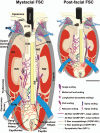
Schematic illustration of representative mystacial and post-facial hyrax follicle-sinus complexes (FSCs) characterizing innervation types and sensory nerve endings observed. Labels for mystacial FSC also apply to post-facial FSC. Each is characterized by a dense connective tissue capsule, dense innervation (particularly Merkel ending complexes), a circumferential ring sinus, a prominent ringwulst, and a dense mesenchymal sheath. The vibrissa of the mystacial FSC (but not the postfacial FSC) exhibited a ridged appearance at the level of the lower ring sinus continuing through the majority of the cavernous sinus. The relative scale of each FSC is approximately accurate, but innervation is disproportionately scaled to optimize visualization. Scale bar = 1mm. BM = basement membrane, DVN = deep vibrissal nerve, ICB = inner conical body, IRS = inner root sheath, MB = mesenchymal bulge, MS = mesenchymal sheath, OCB = outer conical body, ORS = outer root sheath, RRC = rete ridge collar, RS = ring sinus, RW = ringwulst, SG = sebaceous gland, SVN = superficial vibrissal nerve, VEN = venous supply.
Figure 3
Figure 3
Longitudinal sections just off of the central axis of rock hyrax FSCs illustrating the overall structure, prominent ring sinus and ringwulst, and types of nerve endings present in facial and post-facial body regions. A) mystacial, B) submental, C) lateral body, and D) ventral body region FSCs exhibit the characteristics of true vibrissae. Arrowheads indicate mesenchymal bulges at the ring sinus level. Immunolabeling shown consisted of DAPI (blue, nuclear marker; 4',6-diamidino-2-phenylindole), NF200 (red; 200kD subunit of neurofilament), and PGP (green, universal neuronal marker; protein gene product 9.5). Scale bars=300μm (A-D). Cap = capsule, CS = cavernous sinus, Epi = epidermis, FNEs = free nerve endings, HP = hair papilla, ICB = inner conical body, MEs = Merkel endings, OCB = outer conical body, RRC = rete ridge collar, RS = ring sinus, RW = ringwulst, SG = sebaceous gland, TRB = trabeculae.
Figure 4
Figure 4
Representative innervation of rock hyrax FSCs at the dermal, rete ridge collar, outer and inner conical body levels. A-B) Innervation associated with upper dermal arteries included sympathetic innervation (NPY+, indicated by arrows) and C-fiber innervation (CGRP+, indicated by arrowheads). C) Innervation at the rete ridge collar (RRC) and epidermal (Epi) level (representative mystacial follicle-sinus complex shown), including bilateral bulges of Merkel ending complexes (MECs) seen as terminations of Aβ fibers from the superficial vibrissal nerve (SVN; seen in more detail in D). Prominent sebaceous glands (SGs) were also observed. E) In addition to MECs, representative innervation at the RRC and inner conical body (ICB) levels included fine-caliber innervation consisting of C- and Aδ-fibers (NF200− and NF200+, respectively) terminating as presumptive free nerve endings (FNEs) in a mystacial follicle-sinus complex. Scale bars=150μm. Epi = epidermis, FNE = free nerve ending, ICB = inner conical body, MEC = Merkel ending complexes, RRC = rete ridge collar, SG = sebaceous gland, SVN = superficial vibrissal nerve. Immunolabeling shown consisted of NPY (red; neuropeptide Y, sympathetic innervation marker) paired with CGRP (green; calcitonin gene-related peptide) (A-B); or CGRP (red) or NF200 (red; 200kD subunit of neurofilament) paired with PGP (green, universal neuronal marker; protein gene product 9.5) (C-E).
Figure 5
Figure 5
Confocal surface reconstructions illustrating the three-dimensional structure of representative hyrax follicle-sinus complex (FSC) innervation and mechanoreceptors. A) Epidermal and rete ridge collar (RRC) innervation, including small-caliber fibers and Merkel endings (submental FSC). B) Small-caliber innervation at the level of the inner conical body (mystacial FSC). C-D) Semicircular cusp of Merkel innervation at the level of the rete ridge collar (mystacial FSC). Arrowheads demarcate edges of the Merkel complex cusp. E) Extensive large-caliber innervation and Merkel endings at the level of the ring sinus (mystacial FSC). F) Large-caliber innervation and club endings at the level of the ring sinus (mystacial FSC). Immunolabeling shown consisted of PGP (universal neuronal marker; protein gene product 9.5).
Figure 6
Figure 6
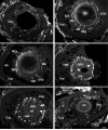
Cross-sectional series from a hyrax mystacial follicle-sinus complex, from superficial to deep levels (A-F). Prominent sebaceous glands are evident (A) and a dense connective tissue capsule surrounds the follicle and its affiliated dense innervation (B-F). Dense Merkel cell innervation can be seen at the ring sinus level surrounding the follicle circumferentially and terminating within the outer root sheath (C). The irregular morphology of the hair follicle becomes apparent at the lower ring sinus and cavernous sinus levels where prominent longitudinal ridges appear (white arrowheads; D-E), and an absence of lateral innervation (within the trabeculae) at the cavernous sinus level is evident (D-F). Immunolabeling shown consisted of PGP (universal neuronal marker; protein gene product 9.5). Scale bar = 300μm (A-F). Cap=capsule, GM=glassy membrane, HP=hair papilla, HS = hair shaft, ICB=inner conical body, MCs=Merkel cells, MS=mesenchymal sheath, OCB=outer conical body, ORS=outer root sheath, RS=ring sinus, RW=ringwulst, SG=sebaceous gland, TRB=trabeculae.
Figure 7
Figure 7
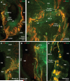
Characterization of hyrax follicle-sinus complex (FSC) innervation at the ring sinus (RS) level (longitudinal planes of section). A) Characteristic innervation at the RS and cavernous sinus (CS) levels of a representative follicle-sinus complex from the caudal body. Arrowhead denotes a mesenchymal bulge at the ring sinus level in proximity to the ringwulst (RW). Large-caliber fibers from the deep vibrissal nerve (DVN) can be seen ascending superficially through the cavernous sinus, curving around the mesenchymal bulge to terminate as Merkel ending complexes (MECs). B) At the level of the ring sinus, club endings and MEC networks can be observed. A mesenchymal bulge at the ring sinus level in proximity to the ringwulst is visible (arrowheads), and presumptive “tangle” endings are apparent at the upper RS/lower ICB level as terminations of large-caliber fibers. C) Fine-caliber innervation is observed at the outer and inner conical body levels, whereas the ring sinus is characterized by dense, circumferential networks of MECs. The mesenchymal bulge proximal to the ringwulst is denoted by the arrowhead. D) Longitudinal lanceolate endings were present along the mesenchymal sheath at the upper extent of the ring sinus, but were relatively sparsely distributed. E) MECs terminate in the outer root sheath of the follicle at the ring sinus level, and large tangle endings were observed. Scale bars=300μm (A-D), 75μm (E). Cap = capsule, CS = cavernous sinus, DVN = deep vibrissal nerve, HP = hair papilla, ICB = inner conical body, LLEs = longitudinal lanceolate endings, MECs = Merkel ending complexes, OCB = outer conical body, RS = ring sinus, RW = ringwulst, TRB = trabeculae.
Figure 8
Figure 8
Characterization of hyrax follicle-sinus complex (FSC) innervation at the cavernous sinus (CS) level, including innervation within the hair papilla (HP) (longitudinal planes of section). A) Spiny ending at the superficial CS level of a dorsal body FSC. B) Submental FSC at the CS level showing spray and reticular endings immunolabeled for NF200. C-E) Limited C- and Aδ-fiber innervation (arrowheads pointing to NF200− labeling and arrows pointing to NF200+ labeling, respectively, in C) within the hair papilla was not superficially extensive (i.e., did not extend very far in the direction of the epidermis). Scale bars=300μm (A), 75μm (B, D), 150μm (C, E). BM = basement membrane.
Figure 9
Figure 9
Hyrax guard hair innervation. In contrast to FSCs, guard hairs lacked a circumferential ring sinus and exhibited only limited density and types of innervation. This innervation was characterized by piloneural complexes that included circumferential longitudinal lanceolate endings (A-B; longitudinal plane of section shown in A, oblique plane of section shown in B). Immunolabeling shown consisted of NF200 (red; 200kD subunit of neurofilament) paired with PGP (green, universal neuronal marker; protein gene product 9.5) or S100 (green, glial and ependymal marker). Scale bars=150μm. Epi = epidermis, HS = hair shaft, LLEs = longitudinal lanceolate endings.

Similar articles

Cited by

  • The functional anatomy of elephant trunk whiskers.
    Deiringer N, Schneeweiß U, Kaufmann LV, Eigen L, Speissegger C, Gerhardt B, Holtze S, Fritsch G, Göritz F, Becker R, Ochs A, Hildebrandt T, Brecht M. Deiringer N, et al. Commun Biol. 2023 Jun 8;6(1):591. doi: 10.1038/s42003-023-04945-5. Commun Biol. 2023. PMID: 37291455 Free PMC article.
  • Constraints on the deformation of the vibrissa within the follicle.
    Luo Y, Bresee CS, Rudnicki JW, Hartmann MJZ. Luo Y, et al. PLoS Comput Biol. 2021 Apr 1;17(4):e1007887. doi: 10.1371/journal.pcbi.1007887. eCollection 2021 Apr. PLoS Comput Biol. 2021. PMID: 33793548 Free PMC article.

References

    1. Ahl AS. The role of vibrissae in behavior: A status review. Vet Res Commun. 1986;10:245–268. - PubMed
    1. Albrecht PJ, Hines S, Eisenberg E, Pud D, Finlay DR, Connolly MK, Pare M, Davar G, Rice FL. Pathologic alterations of cutaneous innervation and vasculature in affected limbs from patients with complex regional pain syndrome. Pain. 2006;120:244–266. - PubMed
    1. Andl T, Reddy ST, Gaddapara T, Millar SE. Wnt signals are required for the initiation of hair follicle development. Dev Cell. 2002;2:643–653. - PubMed
    1. Asher RJ, Novacek MJ, Geisler JH. Relationships of endemic african mammals and their fossil relatives based on morphological and molecular evidence. J Mammal Evol. 2003;10:131–194.
    1. Barry RE, Shoshani J. Heterohyrax brucei. Mammalian species. 2000;645:1–7.

Publication types

Substances

LinkOut - more resources