Deflection of a vibrissa leads to a gradient of strain across mechanoreceptors in a mystacial follicle - PubMed Skip to main page content
U.S. flag

An official website of the United States government

Dot gov

The .gov means it’s official.
Federal government websites often end in .gov or .mil. Before sharing sensitive information, make sure you’re on a federal government site.

Https

The site is secure.
The https:// ensures that you are connecting to the official website and that any information you provide is encrypted and transmitted securely.

Access keys NCBI Homepage MyNCBI Homepage Main Content Main Navigation
. 2015 Jul;114(1):138-45.
doi: 10.1152/jn.00179.2015. Epub 2015 Apr 8.

Deflection of a vibrissa leads to a gradient of strain across mechanoreceptors in a mystacial follicle

Affiliations

Deflection of a vibrissa leads to a gradient of strain across mechanoreceptors in a mystacial follicle

Samuel J Whiteley et al. J Neurophysiol. 2015 Jul.

Abstract

Rodents use their vibrissae to detect and discriminate tactile features during active exploration. The site of mechanical transduction in the vibrissa sensorimotor system is the follicle sinus complex and its associated vibrissa. We study the mechanics within the ring sinus (RS) of the follicle in an ex vivo preparation of the mouse mystacial pad. The sinus region has a relatively dense representation of Merkel mechanoreceptors and longitudinal lanceolate endings. Two-photon laser-scanning microscopy was used to visualize labeled cell nuclei in an ∼ 100-nl vol before and after passive deflection of a vibrissa, which results in localized displacements of the mechanoreceptor cells, primarily in the radial and polar directions about the vibrissa. These displacements are used to compute the strain field across the follicle in response to the deflection. We observe compression in the lower region of the RS, whereas dilation, with lower magnitude, occurs in the upper region, with volumetric strain ΔV/V ∼ 0.01 for a 10° deflection. The extrapolated strain for a 0.1° deflection, the minimum angle that is reported to initiate a spike by primary neurons, corresponds to the minimum strain that activates Piezo2 mechanoreceptor channels.

Keywords: Merkel cells; biomechanics; displacement; ringwulst; somatosensation; whisker.


Figures

Fig. 1.
Fig. 1.
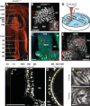
Mouse follicle-sinus complex anatomy and mechanoreceptor distribution. A: anatomical features of the mouse follicle. Merkel cells were selectively labeled with red fluorescent protein (RFP) in AdvillinCre/+ knock-in mice and 100 μm-thick serial sections imaged on a light microscope. RRC, rete ridge collar; OCB, outer conical body; ICB, inner conical body; MDR, Merkel cell-dense region; MS, mesenchymal sheath; RS, ring sinus; RW, ringwulst; DVN, deep vibrissal nerve; IM, intrinsic muscle; CS, cavernous sinus; VS, vibrissa shaft; HB, hair bulb; HP, hair papilla. Directions are: C, caudal; L, lateral. Scale bar, 500 μm. B: maximum projection of a confocal image stack through the MDR of an AdvillinCre/+ knock-in mouse crossed with an RFP reporter mouse. The Merkel cells are located at the level of the RS between the RW and the ICB. Scale bar, 100 μm. Inset (red boxes): magnified view of a single confocal layer close to the edge of the RS, demonstrating that Merkel cells are located in the outer root sheath (ORS). Glassy membrane (GM; unlabeled) is located between the ORS and the MS. Scale bar, 10 μm. C: maximum projection of a confocal image stack of the RS region in a Thy1-TN-XXL transgenic mouse with labeled Merkel and lanceolate-ending afferents. Inset: zoomed-in region of Merkel and lanceolate endings at the level of the MDR. Scale bar, 100 μm. D: schematic of a microdissected follicle row, pinned to a silicone base, immersed in artificial cerebrospinal fluid (aCSF) for 2-photon laser-scanning microscope (TPLSM) imaging. The imaged follicle was suspended above a gap in the silicone base to minimize friction during vibrissa deflection (arrow). E: maximum projection of a TPLSM-acquired image stack of a 4′,6-diamidino-2-phenylindole (DAPI)-labeled vibrissa follicle. A window was opened in the vibrissa capsule above the dorsal aspect of the RS to expose the region between the RW and the ICB. Scale bar, 100 μm. Inset: zoomed-in region containing horizontally elongated cell nuclei that were classified as putative Merkel cells. Scale bar, 10 μm. F: longitudinal cross-section through the image stack in E. Layers of tissue were identified based on DAPI labeling: the MS, GM, and ORS. The inner root sheath (IRS) was never labeled by DAPI. Directions are: R, rostral; M, medial. Yellow lines denote boundaries between the layers labeled above image. Scale bar, 100 μm. G: radial cross-section through the image stack in E, demonstrating the same DAPI-labeled layers as in F. Scale bar, 20 μm. H and I: fixed and sectioned follicle tissue labeled with the membrane dye 5-hexadecanoylamino-fluorescein. The mouse was perfused and fixed, while the vibrissa was deflected in the caudal (H) or rostral (I) directions. Note how the VS buckles and bends in the region of the CS only during rostral deflections. Black arrows indicate the intrinsic sling muscle. Scale bars, 500 μm.
Fig. 2.
Fig. 2.
Measuring relative displacements and strain within the follicle-sinus complex. A: cartoon of the vibrissa in a follicle-sinus complex. The MDR under study is in red and green. B, top: maximum (MAX) projection of the raw image stacks acquired with the vibrissa in its reference (rest) position (red) and when deflected 10° in the caudal direction (green). Inset: magnified image of the boxed region in white, demonstrating rigid rotation and translation of individual DAPI-labeled cell nuclei (yellow denotes overlap). Bottom: maximum projections of radial sections along the longitudinal direction of the follicle. V, ventral direction. C, top: maximum projection of the same image stacks after rigid alignment of the deflected stack onto the reference stack through a rigid transformation with 6° of freedom. Inset: magnified region (same as in A), demonstrating remaining relative movements that cannot be corrected by the transformation (red or green pixels). Bottom: maximum projections of radial sections along the longitudinal direction of the follicle. Scale bars (B and C), 100 μm. D: distribution of displacement vector magnitudes of individual pixels from a single vibrissa deflection, computed from 3-dimensional cross-correlations between aligned image stacks (see methods). RMS, root-mean-squared. E: the coordinate system of pixel-displacement vectors. Each pixel was displaced in 3 directions in a vibrissa-oriented coordinate system: radial (Δr) perpendicular to the VS (purple cylinder), polar (Δα) about the axis of the vibrissa (red arc), and longitudinal (Δl) along the axis of the vibrissa. DAPI-labeled cells included in the analysis were all located within a 90- to 100-μm-thick annulus, approximately bound by the MS and ORS (green cylinder with single planar imposed on the front surface; gray cells are outside of the included volume). F: aligned and transformed image of DAPI-labeled pixels included in analysis of displacement and strain fields in a single experiment. The approximate extents of the MDR and RW are indicated. Gray pixels indicate pixels in which none or insufficient data were available to compute displacements. G: displacement analysis of 10° vibrissa deflection in the caudal direction in a single experiment. Displacements of individual pixels were transformed from Cartesian coordinates into cylindrical coordinates, and then displacements in the radial, polar, and longitudinal directions were averaged across pixels in the radial direction. Note that all displacement maps extend from approximately −π/2 to π/2, which corresponds to the caudal and rostral aspects of the follicle, respectively. Left: radial displacements, Δr, with positive (red/yellow) and negative (blue/white) corresponding to outward and inward displacements, respectively. Middle: polar displacements, Δα, with positive and negative values indicating anterior and posterior rotation over the dorsal side of the VS, respectively. Right: longitudinal displacements, Δl, with positive and negative values indicating inward (away from skin) and outward (toward skin) motion along the axis of the vibrissa, respectively. Displacement field averages were smoothed with a square boxcar mean filter (20 μm width/height). Scale bars (F and G), 100 μm. Insets: SE computed across repetitions of the same deflection (n = 3 trials).
Fig. 3.
Fig. 3.
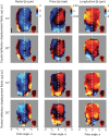
Population analysis of displacement fields. Radial, polar, and longitudinal displacement fields were averaged across follicles by vibrissa deflection direction, i.e., caudal or rostral and amplitude (10° or 20°). The number of deflection conditions varied among experiments. Thus the number of follicles included in each panel was as follows: caudal 10° (n = 7), caudal 20° (n = 4), rostral 10° (n = 5), and rostral 20° (n = 4). Displacement field averages were smoothed by a square mean filter (20 μm width/height). Vertical, dashed lines indicate the axis of the vibrissae. Scale bar, 100 μm.
Fig. 4.
Fig. 4.
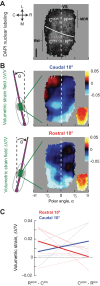
Population analysis of strain fields. A: raw DAPI fluorescence image aligned and averaged across follicles and then transformed into cylindrical coordinates (see methods). The boundary between the RW and MDR is indicated by the curved, solid white line. Vertical, dashed line indicates the center axis of the VS. Scale bar, 100 μm. CMDR, caudal MDR; RMDR, rostral MDR; CRW, caudal RW; RRW, rostral RW. B: strain fields were averaged across follicles by vibrissa deflection direction (caudal or rostral). The number of follicles included in each panel is as in Fig. 3. Strain field averages were smoothed by a square median filter across 100 μm. The cartoons indicate the direction of deflection and the area (green) for which volume strains were computed (ΔV/V; see text). C: gradients of mean strain across diagonal quadrants in the follicle. Dashed lines are individual follicles, and solid lines are averages.

Similar articles

Cited by

References

    1. Abraira VE, Ginty DD. The sensory neurons of touch. Neuron 79: 618–639, 2013. - PMC - PubMed
    1. Bagdasarian K, Szwed M, Knutsen PM, Deutsch D, Derdikman D, Pietr M, Simony E, Ahissar E. Pre-neuronal morphological processing of object location by individual whiskers. Nat Neurosci 16: 622–631, 2013. - PubMed
    1. Coste B, Mathur J, Schmidt M, Earley T. Piezo1 and Piezo2 are essential components of distinct mechanically activated cation channels. Science 330: 55–60, 2010. - PMC - PubMed
    1. da Silva S, Hasegawa H, Scott A, Zhou X, Wagner AK, Han BX, Wang F. Proper formation of whisker barrelettes requires periphery-derived Smad4-dependent TGF-beta signaling. Proc Natl Acad Sci USA 22: 3395–3400, 2011. - PMC - PubMed
    1. Ebara S, Kumamoto K, Matsuura T, Mazurkiewicz JE, Rice FL. Similarities and differences in the innervation of mystacial vibrissal follicle-sinus complexes in the rat and cat: a confocal microscopic study. J Comp Neurol 449: 103–119, 2002. - PubMed

Publication types

LinkOut - more resources