Arrhythmogenesis in a catecholaminergic polymorphic ventricular tachycardia mutation that depresses ryanodine receptor function - PubMed Skip to main page content
U.S. flag

An official website of the United States government

Dot gov

The .gov means it’s official.
Federal government websites often end in .gov or .mil. Before sharing sensitive information, make sure you’re on a federal government site.

Https

The site is secure.
The https:// ensures that you are connecting to the official website and that any information you provide is encrypted and transmitted securely.

Access keys NCBI Homepage MyNCBI Homepage Main Content Main Navigation
. 2015 Mar 31;112(13):E1669-77.
doi: 10.1073/pnas.1419795112. Epub 2015 Mar 16.

Arrhythmogenesis in a catecholaminergic polymorphic ventricular tachycardia mutation that depresses ryanodine receptor function

Affiliations

Arrhythmogenesis in a catecholaminergic polymorphic ventricular tachycardia mutation that depresses ryanodine receptor function

Yan-Ting Zhao et al. Proc Natl Acad Sci U S A. .

Abstract

Current mechanisms of arrhythmogenesis in catecholaminergic polymorphic ventricular tachycardia (CPVT) require spontaneous Ca(2+) release via cardiac ryanodine receptor (RyR2) channels affected by gain-of-function mutations. Hence, hyperactive RyR2 channels eager to release Ca(2+) on their own appear as essential components of this arrhythmogenic scheme. This mechanism, therefore, appears inadequate to explain lethal arrhythmias in patients harboring RyR2 channels destabilized by loss-of-function mutations. We aimed to elucidate arrhythmia mechanisms in a RyR2-linked CPVT mutation (RyR2-A4860G) that depresses channel activity. Recombinant RyR2-A4860G protein was expressed equally as wild type (WT) RyR2, but channel activity was dramatically inhibited, as inferred by [(3)H]ryanodine binding and single channel recordings. Mice heterozygous for the RyR2-A4860G mutation (RyR2-A4860G(+/-)) exhibited basal bradycardia but no cardiac structural alterations; in contrast, no homozygotes were detected at birth, suggesting a lethal phenotype. Sympathetic stimulation elicited malignant arrhythmias in RyR2-A4860G(+/-) hearts, recapitulating the phenotype originally described in a human patient with the same mutation. In isoproterenol-stimulated ventricular myocytes, the RyR2-A4860G mutation decreased the peak of Ca(2+) release during systole, gradually overloading the sarcoplasmic reticulum with Ca(2+). The resultant Ca(2+) overload then randomly caused bursts of prolonged Ca(2+) release, activating electrogenic Na(+)-Ca(2+) exchanger activity and triggering early afterdepolarizations. The RyR2-A4860G mutation reveals novel pathways by which RyR2 channels engage sarcolemmal currents to produce life-threatening arrhythmias.

Keywords: CPVT; cardiac arrhythmias; heart; ryanodine receptor; sarcoplasmic reticulum.


Conflict of interest statement

The authors declare no conflict of interest.

Figures
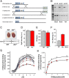
Fig. 1.
Fig. 1.
Generation of mice harboring the RyR2-A4860G mutation and functional assessment of their RyR2 activity. (A) Strategy for generation of the knock-in mice by homologous recombination. Lines a and b show minitargeting vector and retrieval vector generated the targeting vector (line c) by homologous recombination (crosses). Line c shows the RYR2 targeting vector containing the A4860G mutation and the loxP flanked NEO cassette and the HSV-TK cassette. Line d shows the homologous recombination (crosses) between the endogenous RyR2 locus and the A4860G knock-in targeting vector. Line e shows the targeted chromosome 13 containing the 4860G mutation. Line f shows the floxed allele after Cre excision of the Neo cassette. (B) PCR confirmation of WT and heterozygous mice. The absence of the 620-bp (Upper) indicates the mutated allele. (C) Whole hearts of WT and RyR2-A4860G+/− heterozygous mice are structurally indistinguishable by gross appearance and echocardiographic parameters. (Bottom) Western blot of five different animals from each group. (D) The density of the RyR2 protein was similar for both groups. (E) Heart rate was measured in anesthetized animals (n = 8 for each group) during echocardiography. (F) Non-Mendelian propagation of RyR2-A4860G heterozygous mice yields ∼30% WT, ∼70% heterozygotes, and 0% homozygotes. (G) [3H]Ryanodine binding to whole heart homogenates of WT (gray symbols) or RyR2-A4860G+/− (red symbols) mice yield variable binding activity in the latter group only. (H) [3H]Ryanodine binding isotherms for pooled WT (gray symbols) and RyR2-A4860G+/− (red symbols) heart homogenates under conditions that maximize RyR2 activity. *P < 0.05 vs. WT.
Fig. 2.
Fig. 2.
Assessment of e–c coupling gain in WT and RyR2-A4860G+/− ventricular myocytes. (A) Representative ICaL (Top), laser scanning confocal Ca2+ image (Middle), and fluorescence intensity plot of the Ca2+ image (Bottom) from a stimulation step to 0 mV. (B) I–V plot of ICaL density (pA/pF) was similar for WT (black symbols) and RyR2-A4860G+/− (red symbols) cells. Rise rate (C), as well as amplitude (D) of the intracellular Ca2+ transient, was lower for RyR2-A4860G+/− cells (*P < 0.05, **P ≤ 0.01, and ***P ≤ 0.001). (E) e–c coupling gain, calculated as the ratio of [Ca2+]i transient amplitude (ΔF/F0) vs. ICaL density (pA/pF), was significantly lower for RyR2-A4860G+/− cells, especially at positive test potentials (n = 10 cells from n = 8 mice for each group).
Fig. 3.
Fig. 3.
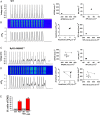
EADs and anomalous [Ca2+]i transients in RyR2-A4860G+/− ventricular myocytes stimulated with ISO. Representative APs of WT (A) and RyR2-A4860G+/− (C) cells and their associated [Ca2+]i transients (B and D). Poincaré plots show uniform APD90 and resting potential (A, Middle and Right, respectively) between stimulating pulses and uniform amplitude and decay time for their associated [Ca2+]i transients (B, Middle and Right, respectively) for WT cells, but variability in the same parameters (C and D, Middle and Right, respectively) for RyR2-A4860G+/− cells. (E) WT ventricular myocytes rarely exhibited EADs during the stimulation protocol, but they were frequent in RyR2-A4860G+/− cells, especially after ISO stimulation. Incidence of EADs: only 1 in 13 (no ISO) and 1 in 10 (with ISO) WT cells displayed EADs, whereas 7 in 15 (no ISO) and 8 in 13 (with ISO) RyR2-A4860G+/− cells showed EADs and abnormal Ca2+ release (*P < 0.05 vs. WT; #P < 0.05 vs. WT with ISO; WT, n = 6 mice; heterozygous, n = 8 mice).
Fig. 4.
Fig. 4.
SR Ca2+ load in RyR2-A4860G+/− cells decreases after EADs. SR Ca2+ load (estimated by measuring the peak of the caffeine-induced Ca2+ release) was higher in RyR2-A4860G+/− cells that did not exhibit EADs during the stimulation protocol (A) than in those exhibiting EADs during identical protocols (B). Bars in C represent the SR Ca2+ load from a group of 30 WT cells, 28 RyR2-A4860G+/− cells without EADs, and 11 RyR2-A4860G+/− cells with EADs. A total of 300 nM ISO was applied in all cells (WT, n = 5 mice; heterozygous, n = 7 mice). **P < 0.01 vs. WT; *P < 0.05 between the two RyR2-A4860G+/− groups.
Fig. 5.
Fig. 5.
Inhibition of the sarcolemmal NCX abolishes EADs in RyR2-A4860G+/− cells. (A) Ventricular myocyte APs during [Ca2+]i transients with prolonged phase of Ca2+ release before (black line) and after (red line) perfusion of 300 nM CB-DMB. Cells were stimulated with 300 nM ISO for at least 15 min before patch-clamping. Resting potential (B) and AP amplitude (C) were not altered by CB-DMB (300 nM) treatment, but APD (D) substantially decreased. (E) The percentage of cells displaying EADs decreased with CB-DMB treatment, from 8 of 14 (57%) to 3 of 11 (27%) to 0 of 2 (0%) after 0, 300 nM, and 3 µM CB-DMB, respectively. Data are from six mice.
Fig. 6.
Fig. 6.
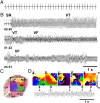
Optical mapping reveals VF in RyR2-A4860G+/− hearts. Volume-conducted ECG from a RyR2-A4860G+/− heart at sinus baseline (A) and after perfusion with 200 nM ISO and 3.6 mM CaCl2 (B). SR, sinus rhythm; VT, ventricular tachycardia. Elapsed time (min) is indicated underneath the traces. (C) Phase map of a rotor on the anterior ventricular epicardial surface superimposed on a digital snapshot of a mouse heart. (D) Sequential 2D phase maps of meandering rotors maintaining VF in a RyR2-A4860G+/− heart. Colors indicate progressive increase in phase over space covering a complete cycle from −π to +π. All phases (colors) converge on a phase singularity (rotor) near the center of each map. Curved arrows indicate sense of rotation. Vertical arrows on ECG trace (Bottom) indicate the time points corresponding to phase maps ae. LA, left atrium; LV, left ventricle; RA, right atrium; RV, right ventricle.
Fig. 7.
Fig. 7.
Hypothetical mechanisms generating EADs in RyR2-A4860G+/− ventricular myocytes. In a single RyR2-A4860G+/− cardiomyocyte containing functionally divergent RyR2 channels (WT shown in violet, AG shown in bronze), a normal ICaL first elicits a [Ca2+]i transient that displays a single phase of Ca2+ release and turns off normally, although its amplitude is lower than that of WT cells (first and second pulses). The lower amplitude of the [Ca2+]i transient despite normal ICaL (decreased e–c coupling gain) leaves a residual amount of Ca2+ inside the SR, which gradually builds up. This process is repeated n times until the SR Ca2+ load reaches a threshold that is sufficient to activate hyporesponsive RyR2 channels, which then release Ca2+ and further activate neighboring RyR2 channels, enhancing CICR. The activation of hyporesponsive RyR2 channels immediately after an initial burst of Ca2+ release (peak) generates a second, protracted and low-amplitude phase of Ca2+ release (pedestal) that supercharges the sarcolemmal NCX, generating in turn an Iti that depolarizes the membrane further, creating an EAD. If SR Ca2+ release during the pedestal returns SR load to below threshold, a new equilibrium ensues, and EADs will be absent until SR load increases again.

Similar articles

Cited by

References

    1. Bers DM. Calcium cycling and signaling in cardiac myocytes. Annu Rev Physiol. 2008;70:23–49. - PubMed
    1. Van Petegem F. Ryanodine receptors: Structure and function. J Biol Chem. 2012;287(38):31624–31632. - PMC - PubMed
    1. Capes EM, Loaiza R, Valdivia HH. Ryanodine receptors. Skelet Muscle. 2011;1(1):18. - PMC - PubMed
    1. Xie Y, Grandi E, Puglisi JL, Sato D, Bers DM. β-adrenergic stimulation activates early afterdepolarizations transiently via kinetic mismatch of PKA targets. J Mol Cell Cardiol. 2013;58:153–161. - PMC - PubMed
    1. Priori SG, et al. Mutations in the cardiac ryanodine receptor gene (hRyR2) underlie catecholaminergic polymorphic ventricular tachycardia. Circulation. 2001;103(2):196–200. - PubMed

Publication types

Supplementary concepts

LinkOut - more resources