Innervation patterns of sea otter (Enhydra lutris) mystacial follicle-sinus complexes
- PMID: 25400554
- PMCID: PMC4212681
- DOI: 10.3389/fnana.2014.00121
Innervation patterns of sea otter (Enhydra lutris) mystacial follicle-sinus complexes
Abstract
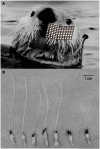
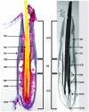
Sea otters (Enhydra lutris) are the most recent group of mammals to return to the sea, and may exemplify divergent somatosensory tactile systems among mammals. Therefore, we quantified the mystacial vibrissal array of sea otters and histologically processed follicle-sinus complexes (F - SCs) to test the hypotheses that the number of myelinated axons per F - SC is greater than that found for terrestrial mammalian vibrissae and that their organization and microstructure converge with those of pinniped vibrissae. A mean of 120.5 vibrissae were arranged rostrally on a broad, blunt muzzle in 7-8 rows and 9-13 columns. The F-SCs of sea otters are tripartite in their organization and similar in microstructure to pinnipeds rather than terrestrial species. Each F-SC was innervated by a mean 1339 ± 408.3 axons. Innervation to the entire mystacial vibrissal array was estimated at 161,313 axons. Our data support the hypothesis that the disproportionate expansion of the coronal gyrus in somatosensory cortex of sea otters is related to the high innervation investment of the mystacial vibrissal array, and that quantifying innervation investment is a good proxy for tactile sensitivity. We predict that the tactile performance of sea otter mystacial vibrissae is comparable to that of harbor seals, sea lions and walruses.
Keywords: F-SCs; axon investment; comparative neurobiology; marine mammals; otters; peripheral nervous system; somatosensory system; vibrissae.
Figures


Similar articles
-
Innervation patterns of mystacial vibrissae support active touch behaviors in California sea lions (Zalophus californianus).J Morphol. 2019 Nov;280(11):1617-1627. doi: 10.1002/jmor.21053. Epub 2019 Aug 19. J Morphol. 2019. PMID: 31424610
-
Does Vibrissal Innervation Patterns and Investment Predict Hydrodynamic Trail Following Behavior of Harbor Seals (Phoca vitulina)?Anat Rec (Hoboken). 2019 Oct;302(10):1837-1845. doi: 10.1002/ar.24134. Epub 2019 May 3. Anat Rec (Hoboken). 2019. PMID: 30980470
-
Follicle Microstructure and Innervation Vary between Pinniped Micro- and Macrovibrissae.Brain Behav Evol. 2016;88(1):43-58. doi: 10.1159/000447551. Epub 2016 Aug 23. Brain Behav Evol. 2016. PMID: 27548103
-
Microstructure and innervation of the mystacial vibrissal follicle-sinus complex in bearded seals, Erignathus barbatus (Pinnipedia: Phocidae).Anat Rec A Discov Mol Cell Evol Biol. 2006 Jan;288(1):13-25. doi: 10.1002/ar.a.20273. Anat Rec A Discov Mol Cell Evol Biol. 2006. PMID: 16342212
-
Active touch in sea otters: in-air and underwater texture discrimination thresholds and behavioral strategies for paws and vibrissae.J Exp Biol. 2018 Sep 17;221(Pt 18):jeb181347. doi: 10.1242/jeb.181347. J Exp Biol. 2018. PMID: 30224372
Cited by
-
Fossil brains provide evidence of underwater feeding in early seals.Commun Biol. 2023 Aug 17;6(1):747. doi: 10.1038/s42003-023-05135-z. Commun Biol. 2023. PMID: 37591929 Free PMC article.
-
Constraints on the deformation of the vibrissa within the follicle.PLoS Comput Biol. 2021 Apr 1;17(4):e1007887. doi: 10.1371/journal.pcbi.1007887. eCollection 2021 Apr. PLoS Comput Biol. 2021. PMID: 33793548 Free PMC article.
-
Knowing when to stick: touch receptors found in the remora adhesive disc.R Soc Open Sci. 2020 Jan 15;7(1):190990. doi: 10.1098/rsos.190990. eCollection 2020 Jan. R Soc Open Sci. 2020. PMID: 32218935 Free PMC article.
References
-
- Armed Forces Institute of Pathology . (1968). Manual of Histologic Staining Methods. Washington, DC: American Registry of Pathology
-
- Armed Forces Institute of Pathology . (1994). Laboratory Methods in Histotechnology. Washington, DC: American Registry of Pathology
-
- Berta A., Sumich J. L. (1999). Marine Mammals: Evolutionary Biology. San Diego: Academic Press
LinkOut - more resources
Full Text Sources
Other Literature Sources

