Compressive osseointegration promotes viable bone at the endoprosthetic interface: retrieval study of Compress implants
- PMID: 17576554
- PMCID: PMC2551719
- DOI: 10.1007/s00264-007-0392-z
Compressive osseointegration promotes viable bone at the endoprosthetic interface: retrieval study of Compress implants
Abstract
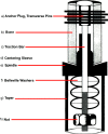
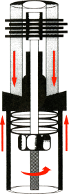
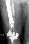
The Compress implant (Biomet, Warsaw, IN) is an innovative device developed to enable massive endoprosthetic fixation through the application of compressive forces at the bone-implant interface. This design provides immediate, stable anchorage and helps to avoid the long-term complication of aseptic loosening secondary to stress shielding and particle-induced osteolysis seen in conventional, stemmed megaprostheses. The purpose of our study was to evaluate the in vivo biological effects of the high compressive forces attained. Twelve consecutive Compress patients undergoing revision surgery for infection, periprosthetic fracture, or local tumour recurrence were reviewed in order to exclude the possibility of osteonecrosis at the prosthetic interface. Compressive forces ranged from 400-800 lb. Duration of implantation averaged 3.3 years (range 0.4-12.2 years). Two patients with infection demonstrated loosening at the bone-prosthetic interface; otherwise, there was no radiographic evidence of prosthetic failure in any of the patients. No patient demonstrated histological evidence of osteonecrosis. In fact, new woven bone and other findings consistent with viable bone were noted in all of the retrieved specimens.
La prothèse Compress® (Biomet, Warsaw, In) est une endo-prothèse massive, innovante, développée pour permettre une fixation avec des forces de compression au niveau des interfaces os-implant. Le dessin de l’implant permet une stabilité immédiate au niveau de l’ancrage et semble éviter des complications, à long terme, comme le descellement aseptique, secondaire à un stress shielding rencontré de façon habituelle dans les méga prothèses. Le but de cette étude est d’évaluer les effets biologiques in vivo de ces forces de compression. 11 prothèses consécutives de type Compress® ont été réalisées chez 11 patients, nécessitant une réintervention pour infection, pour fracture périprothétique ou pour récidive d’une tumeur locale. Les forces de compression ont été évaluées de 400 à 800 lb. Le temps d’implantation moyen a été de 2.5 ans (0.4 à 6.5 ans). Deux patients ont présenté un descellement infectieux à l’interface os-prothèse, il n’a pas été mis en évidence, sur le plan radiographique d’échecs de cet implant chez aucun des patients. Aucun patient n’a également montré de façon évidente des phénomènes d’ostéonécrose histologique et, l’analyse des prothèses explantées a montré qu’il existait au contact de celles-ci un os vivant.
Figures


Similar articles
-
What Are the Long-term Surgical Outcomes of Compressive Endoprosthetic Osseointegration of the Femur with a Minimum 10-year Follow-up Period?Clin Orthop Relat Res. 2022 Mar 1;480(3):539-548. doi: 10.1097/CORR.0000000000001979. Clin Orthop Relat Res. 2022. PMID: 34559734 Free PMC article.
-
Use of Compressive Osseointegration Endoprostheses for Massive Bone Loss From Tumor and Failed Arthroplasty: A Viable Option in the Upper Extremity.Clin Orthop Relat Res. 2017 Jun;475(6):1702-1711. doi: 10.1007/s11999-017-5258-0. Epub 2017 Feb 13. Clin Orthop Relat Res. 2017. PMID: 28194713 Free PMC article.
-
What are the 5-year survivorship outcomes of compressive endoprosthetic osseointegration fixation of the femur?Clin Orthop Relat Res. 2015 Mar;473(3):883-90. doi: 10.1007/s11999-014-3724-5. Clin Orthop Relat Res. 2015. PMID: 24942962 Free PMC article.
-
Limb-sparing surgery for distal femoral and proximal tibial bone lesions: imaging findings with intraoperative correlation.AJR Am J Roentgenol. 2013 Feb;200(2):W193-203. doi: 10.2214/AJR.11.8042. AJR Am J Roentgenol. 2013. PMID: 23345384 Review.
-
Strontium in the bone-implant interface.Dan Med Bull. 2011 May;58(5):B4286. Dan Med Bull. 2011. PMID: 21535993 Review.
Cited by
-
Megaprosthetic reconstruction of the distal femur with a short residual proximal femur following bone tumor resection: a systematic review.J Orthop Surg Res. 2023 Jan 27;18(1):68. doi: 10.1186/s13018-023-03553-7. J Orthop Surg Res. 2023. PMID: 36707881 Free PMC article.
-
The multidisciplinary management of osteosarcoma.Curr Treat Options Oncol. 2009 Apr;10(1-2):82-93. doi: 10.1007/s11864-009-0087-3. Epub 2009 Feb 24. Curr Treat Options Oncol. 2009. PMID: 19238553 Review.
-
Compressive osseointegration of tibial implants in primary cancer reconstruction.Clin Orthop Relat Res. 2009 Nov;467(11):2807-12. doi: 10.1007/s11999-009-0986-4. Epub 2009 Aug 4. Clin Orthop Relat Res. 2009. PMID: 19653050 Free PMC article.
-
Hip-preserving reconstruction using a customized cemented femoral endoprosthesis with a curved stem in patients with short proximal femur segments: Mid-term follow-up outcomes.Front Surg. 2022 Sep 22;9:991168. doi: 10.3389/fsurg.2022.991168. eCollection 2022. Front Surg. 2022. PMID: 36338613 Free PMC article.
-
Osseointegrated Transcutaneous Device for Amputees: A Pilot Large Animal Model.Adv Orthop. 2018 Sep 13;2018:4625967. doi: 10.1155/2018/4625967. eCollection 2018. Adv Orthop. 2018. PMID: 30302292 Free PMC article.
References
-
- Avedian R, Goldsby RE, Kramer MJ, O’Donnell RJ (2007) Effect of chemotherapy on initial compressive osseointegration of oncologic endoprostheses. Clin Orthop Relat Res, Epub March 15 - PubMed
-
- Bini SA, Johnston JO, Martin DL. Compliant prestress fixation in tumor prostheses: interface retrieval data. Orthopedics. 2000;23(7):707–712. - PubMed
-
- Lanyon LE, Hampson WGJ, Goodship AE, Shah JS. Bone deformation recorded in vivo from strain gauges attached to the human tibial shaft. Acta Orthop Scand. 1975;46:256–268. - PubMed
MeSH terms
LinkOut - more resources
Full Text Sources
Medical

