Sub-diffraction-limit imaging by stochastic optical reconstruction microscopy (STORM)
- PMID: 16896339
- PMCID: PMC2700296
- DOI: 10.1038/nmeth929
Sub-diffraction-limit imaging by stochastic optical reconstruction microscopy (STORM)
Abstract
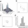
We have developed a high-resolution fluorescence microscopy method based on high-accuracy localization of photoswitchable fluorophores. In each imaging cycle, only a fraction of the fluorophores were turned on, allowing their positions to be determined with nanometer accuracy. The fluorophore positions obtained from a series of imaging cycles were used to reconstruct the overall image. We demonstrated an imaging resolution of 20 nm. This technique can, in principle, reach molecular-scale resolution.
Figures


Comment in
-
Single-molecule mountains yield nanoscale cell images.Nat Methods. 2006 Oct;3(10):781-2. doi: 10.1038/nmeth1006-781. Nat Methods. 2006. PMID: 16990808 Free PMC article.
-
The limits of light.Nat Rev Mol Cell Biol. 2010 Oct;11(10):678. doi: 10.1038/nrm2989. Nat Rev Mol Cell Biol. 2010. PMID: 20861874 No abstract available.
Similar articles
-
Three-dimensional super-resolution imaging by stochastic optical reconstruction microscopy.Science. 2008 Feb 8;319(5864):810-3. doi: 10.1126/science.1153529. Epub 2008 Jan 3. Science. 2008. PMID: 18174397 Free PMC article.
-
Technical review: types of imaging-direct STORM.Anat Rec (Hoboken). 2014 Dec;297(12):2227-31. doi: 10.1002/ar.22960. Epub 2014 Jul 4. Anat Rec (Hoboken). 2014. PMID: 24995970 Review.
-
Super-resolution imaging with stochastic single-molecule localization: concepts, technical developments, and biological applications.Microsc Res Tech. 2014 Jul;77(7):502-9. doi: 10.1002/jemt.22346. Epub 2014 Feb 25. Microsc Res Tech. 2014. PMID: 24616244 Review.
-
Stochastic Optical Reconstruction Microscopy (STORM).Curr Protoc Cytom. 2017 Jul 5;81:12.46.1-12.46.27. doi: 10.1002/cpcy.23. Curr Protoc Cytom. 2017. PMID: 28678417 Free PMC article.
-
A user's guide to localization-based super-resolution fluorescence imaging.Methods Cell Biol. 2013;114:561-92. doi: 10.1016/B978-0-12-407761-4.00024-5. Methods Cell Biol. 2013. PMID: 23931523
Cited by
-
Surpassing the Diffraction Limit in Label-Free Optical Microscopy.ACS Photonics. 2024 Aug 27;11(10):3907-3921. doi: 10.1021/acsphotonics.4c00745. eCollection 2024 Oct 16. ACS Photonics. 2024. PMID: 39429866 Free PMC article. Review.
-
Exploring Lignification Complexity in Plant Cell Walls with Airyscan Super-resolution Microscopy and Bioorthogonal Chemistry.Chem Biomed Imaging. 2023 Jul 6;1(5):479-487. doi: 10.1021/cbmi.3c00052. eCollection 2023 Aug 28. Chem Biomed Imaging. 2023. PMID: 39473934 Free PMC article.
-
Dual-objective STORM reveals three-dimensional filament organization in the actin cytoskeleton.Nat Methods. 2012 Jan 8;9(2):185-8. doi: 10.1038/nmeth.1841. Nat Methods. 2012. PMID: 22231642 Free PMC article.
-
Microscopy beyond the diffraction limit using actively controlled single molecules.J Microsc. 2012 Jun;246(3):213-20. doi: 10.1111/j.1365-2818.2012.03600.x. Epub 2012 Apr 12. J Microsc. 2012. PMID: 22582796 Free PMC article. Review.
-
Dual-color superresolution microscopy reveals nanoscale organization of mechanosensory podosomes.Mol Biol Cell. 2013 Jul;24(13):2112-23. doi: 10.1091/mbc.E12-12-0856. Epub 2013 May 1. Mol Biol Cell. 2013. PMID: 23637461 Free PMC article.
References
Publication types
MeSH terms
Substances
Grants and funding
LinkOut - more resources
Full Text Sources
Other Literature Sources

